 |
Fusion of Red-fluorescent latex bead phagosomes with dextran-loaded lysosomes (green).
Quantitative analysis of phagolysosome fusion can be performed on whole cell lysate (insert) by FACS. |
 |
Intracellular sequestration of MHC class II molecules (green) in macrophages infected with live BCG vaccine (red).
A modified BCG strain that stimulates surface export of Ag-loaded class II molecules would be an attractive novel TB vaccine. |
|
|
Alternative to morbid experimental TB: non-invasive and painless in vivo bioimaging of mice infected with bioluminescent mycobacteria.
Six animals can be used instead of 42 to examine survival to TB infection over a period of 6 months. |

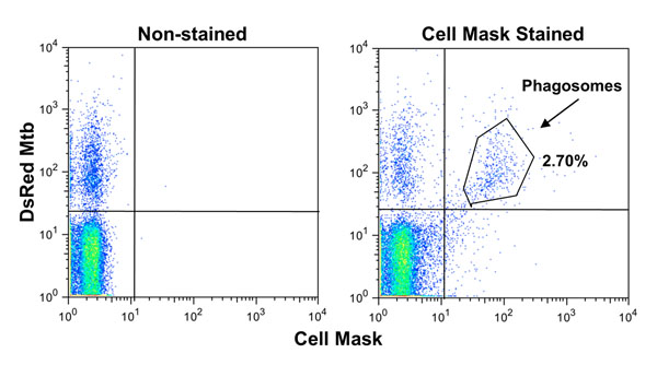
FACS analysis of Mtb phagosomes.
Macrophage cell surface is labeled with CellMask Deep Red (detectable by FL4 channel) prior to infection with DsRed mycobacteria (FL2). Thereafter, cells are homogenized and centrifuged at low speed to remove nuclei and intact cells. The upper fraction corresponds to a crude phagosome preparation in which bacteria are included in cell membrane-derived vacuoles (double FL2/FL4 positive events). Phagosome preparations can be stained with specific antibodies followed by FITC-conjugated secondary antibodies and levels of phagosomal markers (FL1 histograms) can be easily determined by FACS.